Integral System Explained
The purpose of this website is to provide easy-to-understand information about the problems of the pelvic floor to people who are suffering from bladder, bowel, chronic pelvic pain problems.
This website is for women who want to easily find more about their pelvic floor and the health issues, symptoms and available remedies related to this crucially important part of the body.
Historically, pelvic floor dysfunction caused by childbirth and/or due to natural ageing has produced a largely intractable range of debilitating symptoms which can impact enormously on a woman’s quality of life.
We hope that by exploring the information provided here you will not only widen your knowledge of your own body but also discover the new avenues for treatment offered by the Integral System.
On this page below we will educate you about your body and ligaments so you may fully understand the science behind the integral system.
This will be sectioned into three categories:
1) Understanding the Body;
2) Understanding the Ligaments; &
3) Damage and its Consequences
At the end of this page we urge you to learn more about causes, signs and symptoms.
Understanding the Body
Bladder, Bowel and Uterus
The purpose of this website is to provide easy-to-understand information about the problems of the pelvic floor to people who are suffering from bladder, bowel, chronic pelvic pain problems.
This website is for women who want to easily find more about their pelvic floor and the health issues, symptoms and available remedies related to this crucially important part of the body.
Historically, pelvic floor dysfunction caused by childbirth and/or due to natural ageing has produced a largely intractable range of debilitating symptoms which can impact enormously on a woman’s quality of life.
We hope that by exploring the information provided here you will not only widen your knowledge of your own body but also discover the new avenues for treatment offered by the Integral System.
On this page below we will educate you about your body and ligaments so you may fully understand the science behind the integral system.
This will be sectioned into three categories:
1) Understanding the Body;
2) Understanding the Ligaments; &
3) Damage and its Consequences
At the end of section three we urge you to learn more about causes, signs and symptoms. ADD LINK HERE

How the Pelvic Floor Sits in the Body
The Pelvic Muscles
The pelvic muscles (dark red) extend from the tail bone to the pubic bone. They support your vagina, bladder and bowel from below. The red arrows indicate the directions where the muscles contract, backwards to open these organs, forwards to close them.
We saw from the suspension bridge diagram, that the muscles pull against the ligaments. So if your ligaments are loose, the muscle strength weakens, and may not be able to keep your bladder or bowel emptying tubes closed. As a consequence of this, you may feel a leakage of urine, wind or faeces. This is called “incontinence”. Another related condition is failure to close the vaginal tube, so you may feel water entering your vagina during swimming. If your damaged ligaments do not allow your muscles to open these same emptying tubes, you may have to strain to empty your bladder or bowel. This condition is called “evacuation disorder” or “emptying disorder”.
Working Like a Symphony Orchestra
Your vagina, bladder, bowel, muscles, and ligaments are like instruments in an orchestra. The brain is the conductor, and ensures that all the instruments work harmoniously to produce the right music. Every instrument in the orchestra has a specific job. Damage to even one instrument will affect the performance. The brain directs the orchestra to open the bladder (and bowel) or to close it.
Depending on what structure is damaged, the bladder may not be able to close properly, and the patient leaks (“incontinence”); or she may not be able empty her bladder, or she may have both problems. Nerves at the base of the bladder (‘N’ in the diagram which follows) sense when the bladder is full, and send impulses to the brain. These are perceived as urgency, a desire to go to the toilet.
The Brain and its Nerves
A Sophisticated Feedback System
Your brain works like the computer at a big telephone exchange. Think of your nerves as telephone wires going out to the bladder, the vagina and bowel. These organs have sensors which send signals back to the brain via another set of nerves, to inform it as to what is happening. Your brain receives and processes these signals, and depending on what is required, sends out orders via a series of nerves. Most of this co-ordination occurs in “automatic mode”.
You are not aware of what is happening. Sometimes, you can actually instruct the brain. For example, if it is inconvenient for you to empty your bladder or your bowel, you simply pull the muscles upwards to close off the urethral and anal tubes. Sometimes you push down when you wish to assist emptying your urine and faeces. If, during intercourse, you wish to narrow the vagina, you pull the muscles upwards. This action grips the penis, and increases the sensation for both partners.
Understanding the Ligaments & Muscles
The Structure of Ligaments
A ligament is a complicated elastic structure which needs to be both elastic and strong. It relies on its collagen content for strength. Whilst elastin gives ligaments their flexibility, it is collagen which gives them strength.Collagen is what you see in a scar. Collagen fibres work like the steel rods in cement. Single collagen fibres are “glued” together to give ligaments strength, and elastin gives them elasticity.
The Importance of Ligaments
The bladder sits on top of the vagina, and is partly attached to it. Muscles pull against the ligaments to close or open the bladder. Therefore loose ligaments may weaken the muscle contraction to cause problems with closure (incontinence) or opening (passing urine).
A ligament is like a thick cord in a suspension bridge. In fact, the vagina is suspended exactly like a suspension bridge, with the ligaments above, and the muscles (arrows)

The vagina is suspended from above, like a suspension bridge, with the ligaments above, and the muscles (arrows) below. Muscles (arrows) stretch these structures to give them form and strength.
The diagram indicates what your vagina, bladder, and bowel would look like if you had no ligaments to suspend them, a blob of tissue, with no form, no structure, and no strength.
Your organs are suspended from above by the vaginal ligaments – exactly like a suspension bridge. All the ligaments are attached to the vagina and/or uterus. The vagina supports the bladder above, and the rectum (bowel), so anything which damages the vaginal structure, can also affect the bladder and rectum.

Unsuspended ligaments have no shape, strength or function.
The lower part of the uterus is called the cervix, and it is situated right at the back of your vagina. The cervix is where your Pap smear is taken from. Separating the lower end of the vagina from the rectum is a solid mass of tissue called the perineal body. If this is damaged, the rectum (bowel) may bulge forwards into the vagina. This is called a “rectocoele”.
The uterus is an anchoring point for the ligaments – it needs to be preserved if possible.
The role of the uterus in maintaining the function of the pelvic floor is greatly underestimated. Some doctors routinely recommend removal of the uterus during surgery for prolapse. It is preferable to retain the uterus wherever possible, as many important ligaments are attached to it. During the menopause (cessation of the periods), the ovaries cease production of the hormone oestrogen. Since oestrogen is essential for maintaining the strength of the ligaments, the detrimental effects of hysterectomy on prolapse and incontinence become especially evident after the menopause. Hysterectomy (removal of the uterus) reduces the blood supply to the back ligaments, weakening them further. All these factors predispose to prolapse. Prolapse is a general term for bulging of the bladder, uterus or rectum into the vagina.

The uterus functions like the keystone of an arch. Remove the keystone, and the whole Vagina is put at risk of a downward collapse.
Damage and its Consequences
How Ligaments Become Damaged
Pregnancy
Ligaments can be stretched and damaged during Pregnancy and Labour.
As we saw from the suspension bridge diagram above, the muscles pull against the ligaments which support the bridge. If your ligaments are loosened during childbirth, for instance, you may develop a prolapse (a feeling of something coming down), a “dragging” pain low in your abdomen, symptoms in your bladder, for example urgency, frequency, getting up at night, or even problems with bowel emptying or incontinence.
Commencing 6 months before childbirth, the “glue” between the collagen rods begins to soften in response to hormones from the placenta (“afterbirth”). This explains the onset of bladder, bowel, and pain symptoms at this time. Some 24-48 hours before delivery, however, this softening accelerates, and the collagen rods lose 95% of their strength.
During delivery, the baby’s head greatly stretches these collagen rods. Of course, the rods “re-glue” soon after delivery, but often they “re-glue” in a loose and extended position. Neither the ligaments nor the muscles can now work properly, and this may lead to a “fallen womb” (prolapse of the uterus), a bulge of the bladder (cystocoele) or rectum (rectocoele), and a wide range of bladder, bowel and pelvic pain symptoms. Women who have had Caesarian sections may also become incontinent, but less frequently. Loose ligaments may occur in women who have never had children. Such women are born with loose ligaments, or they may have a congenital defect in their collagen.
All these conditions are potentially curable by creation of artificial ligaments, as will be explained later.

The baby’s head stretches and loosens the ligaments and other tissues at childbirth
Age and Menopause
As discussed previously, collagen gives ligaments strength, and elastin gives them elasticity. Both deteriorate markedly after the menopause, and this explains the vast increase in prolapse and incontinence which occurs after this event. A partly damaged ligament which is only just functioning before the menopause, may lose enough collagen after it to “give way”. This may result in organ prolapse, abnormal symptoms, or both.
The oestrogen hormone replacement therapy (“HRT”) helps to slow down the degeneration of collagen. HRT can be applied vaginally, or by patches, creams, tablets, injections. Though oestrogens may be associated with an apparent increased risk of breast cancer, this has to be weighed against their benefits, prevention of osteoporosis, hip fractures, thickening of the vaginal wall in sexually active patients, even perhaps prevention of heart disease. Vaginally inserted oestrogen tablets are considered safe, as they act locally. We recommend these for every post-menopausal woman, even into the most mature age.
Damage and Consequences
As the head descends down through the vagina during childbirth, this may stretch and loosen the ligaments which support the Bladder, Uterus or the Bowel which may cause:
1. Stress incontinence (urine leakage with coughing or exercise).
2. Cystocoele (bulging of the bladder into vagina).
3. Prolapse of the uterus (“fallen womb”).
4. Rectocoele (bulging of the bowel into vagina). All may occur to varying degrees in the same patient.

This diagram illustrates the cystocoele “2”, rectocoele “4”and uterus”3″, all pushing into the vagina, as “lumps”, like a glove turning inside out. All are caused by looseness in the suspensory ligaments and their associated tissues.
Organ Prolapse
A Perspective
The organs bulge to varying degrees. Clearly a severe prolapse, such as the uterine prolapse above, requires treatment. If the bulge is minor, and there are no associated symptoms, there is no need for treatment. However, you may have severe symptoms which may require treatment, even though the prolapse is minor.
Cystocoele
If you held a mirror at your vagina and strained, this is what a cystocoele would look like. The lump “balloons” out from above.
The cause is damage to the middle ligaments and the tissues of the front wall of the vagina, “2”, in the preceding diagrams.

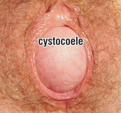
Hover or click above to see image.
A cystocoele bulging out of the vaginal entrance during straining.
Rectocoele
The diagram which follows shows what typical rectocoele looks like. Separating the lower end of the vagina from the rectum is the perineal body.
If this is damaged, the rectum may bulge forwards into the vagina from below.

Rectocoele
Hover or click above to see image.
A rectocoele bulging out of the vagina during straining.
Prolapse of Uterus
The diagram which follows is what a severe prolapse of the uterus (“fallen womb”) looks like.
The cause is damage to the ligaments and the tissues supporting the uterus, “3”, in the preceding diagrams.
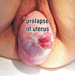
Hover or click above to see image.
A uterus chronically bulging out of the vagina. The white areas are caused by chronic friction.